SPINAL CORD – 3
The image shows part of a cross-section of a spinal cord. It is a higher magnification compared to the figure on the previous page, and its purpose is to identify the gray and white matter of the spinal cord.
The gray matter occupies the center of the spinal cord. It is more intensely stained than the white matter because it contains a large number of neuronal cell bodies and glial cells, as well as a large number of unmyelinated axons and glial cell extensions.
The white matter is less intensely stained and has a reticulated appearance. Most of its fibers are myelinated and are sectioned transversely. Myelin, which covers the axons, is a mixture of complex lipids. Lipids are usually dissolved during the preparation of histological sections by routine techniques, due to the use of ethanol and organic solvents (xylene, benzene, toluene). aS a consequence, places that had myelin now show clear spaces.
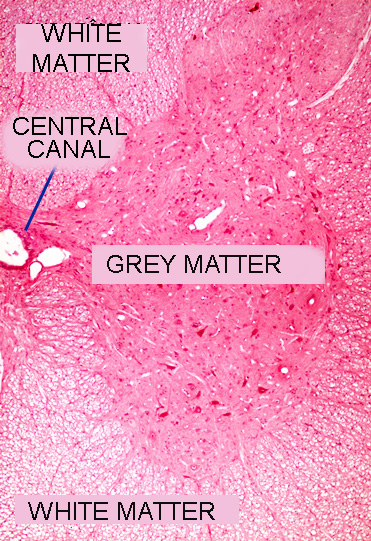
Spinal cord. Staining: HE. Small magnification.
