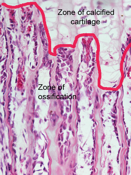
The area above the line is filled with calcified cartilage. It is easy to see that the the space below the line is occupied by another tissue: bone tissue that has been deposited over the calcified cartilage septa.
Lower image
A higher magnification of the transitional area.
In the final region of the calcified cartilage zone, the chondrocytes have undergone cell death, and their nuclei and cytoplasm have disintegrated.
Many sites previously occupied by chondrocytes are now empty (highlighted in green).
In the lower part of the image: walls of cartilage extracellular matrix that surrounded cartilage cells are now covered by a thin layer of bone cells: osteoblasts, osteocytes (highlighted in pink). The newly produced bone matrix is highlighted in a darker tone of blue.
Cartilages do not contain blood vessels. At the center of the image observe a blood vessel (highlighted in brown), that penetrated the cartilage to irrigate the bonethat is being formed.
Epiphyseal disc. Staining: HE. Magnification: small.


Epiphyseal disc. Staining: HE. Magnification: medium.