ARRANGEMENT OF LAMELLAR BONE IN THE DIAPHYSES – 3
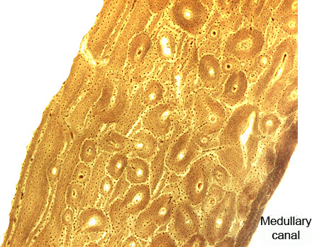
The wall of the diaphysis of mature bones is formed by thousands or millions of very thin bone plates, the bone lamellae. They are made of calcified collagen fibers plus other components of extracellular matrix. Osteocytes are placed between the lamellae.
The wall is composed by three layers of lamellae.
1 – The external or outer circumferential lamellae (highlighted in light green/blue) is placed at the periphery of the diaphysis and made of stacks of slightly curved parallel lamellae.
2 – The thin inner border of the diaphysis, facing the medullary canal, is formed by the internal or inner circumferential lamellae. They are stacks of curved parallel lamellae (highlighted in red).
3 – The widest layer contains the intermediate lamellae, placed between both circumferential lamellae (highlighted in green). It is occupied by Haversian systems and stretches of parallel bone lamellae that fill thr spaces between Haversian systems.
Excepting the articular surfaces, all internal and external surfaces of bones are covered by cell layers called respectively endosteum and periosteum. These layers are formed by bone lining cells (osteoprogenitors plus active and inactive osteoblasts).
The endosteum, usually a thin layer, stays in direct contact with the content of medullary cavity. The thicker periosteum is made of connective tissue proper placed over the osteoprogenitors and osteoblasts.