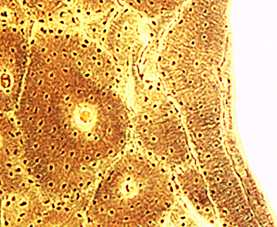
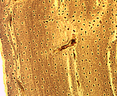
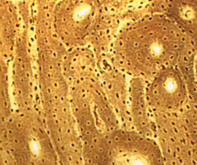
THE ARRANGEMENT OF LAMELLAR BONE IN THE DIAPHYSES – 4
This page discloses the three layers of bone lamellae that form the wall of compact bone of most diaphyses.
The bone sections prepared according to Schmorl’s technique. Images at medium magnification.
1 – Upper image
External circumferential lamellae are a layer of parallel stacks of slightly curved long lamellae. They constitute the outer wall of the diaphysis. This wall is covered by the periosteum, not visible in this type of preparation.
3 – The internal circumferential lamellae layer comprises a series of stacks of slightly curved parallel lamellae that constitute the inner wall of the diaphysis. Its surface is covered by the endosteum, not visible in this type of preparation. The endosteum is in direct contact with the bone marrow. The central bone cavity is to the right of the image.