NON-LAMELLAR BONE AND LAMELLAR BONE – 3
Upper figure – Non-lamellar bone
Osteocytes highlighted in blue are irregularly distributed in the extracellular matrix. Osteoblasts highlighted in light brown. Osteoclast highlighted in red.
Osteocytes highlighted in blue are irregularly distributed in the extracellular matrix. Osteoblasts highlighted in light brown. Osteoclast highlighted in red.
Lower figure – Lamellar bone
Lacunae and canalicles filled with red stain organized in parallel rows. The cabalicles connect each osteocyte with its neighbors.
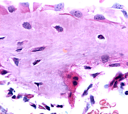
Non-lamellar bone. Staining: HE. Magnification: medium.


Lamellar bone. Thionine stai fills cavities and tunnels placed in the bone matrix. Magnification: small.