NERVES – 2
The Structure of Nerves
Unlike very thin and delicate nerves, larger nerves contain several fascicles of nerve fibers, separated from one another by layers of connective tissue.
The entire nerve is surrounded by an outer layer of dense irregular connective tissue called the epineurium (highlighted in pink). The thickness of the epineurium varies according to the size of the nerve. Nerves located within connective tissue or within an organ may have a less clearly defined epineurium that blends with the surrounding connective tissue.
In medium-sized and large nerves, connective tissue from the epineurium extends into the interior of the nerve, where it surrounds and separates the individual fascicles, as shown in the figure.
The perineurium (highlighted in blue) is a specialized connective tissue layer that surrounds each fascicle of nerve fibers. It contains collagen fibers and cells with elongated nuclei.
The endoneurium (highlighted in red) is a delicate layer of loose connective tissue that surrounds each individual nerve fiber. It contains blood capillaries, collagen fibers, fibroblasts, and small numbers of mast cells and macrophages. Because the endoneurium is very thin and contains only a small amount of connective tissue around each nerve fiber, it may be difficult to distinguish in histological sections.
During tissue processing, the endoneurium may undergo some degree of shrinkage. This can produce artificial spaces between adjacent nerve fibers, which are artifacts of tissue preparation rather than true spaces within the nerve.
Very thin nerves may have only a thin outer connective tissue covering and a small amount of endoneurial connective tissue between their individual nerve fibers.
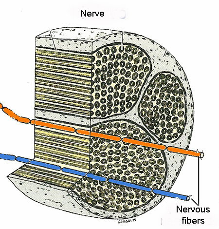
Drawing modified from “Basic Histology”, Junqueira and Carneiro, 12a Ed.
