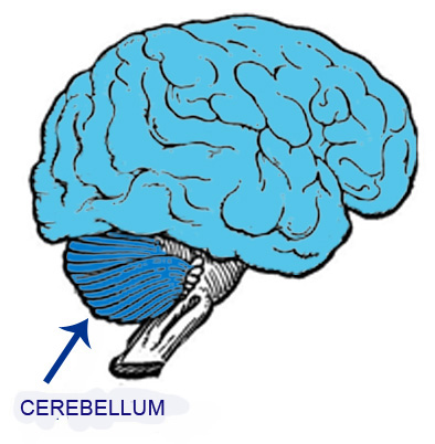
The upper figure is a schematic representation of the brain and cerebellum, which will be examined in greater detail on the following pages.
The central nervous system (CNS) contains regions with a high concentration of neuronal cell bodies and other regions containing large numbers of neuronal processes, particularly axons. These two types of regions can be distinguished macroscopically when we observe a sectione brain or cerebellum because they have different colors.
Regions containing a high concentration of neuronal cell bodies have a grayish appearance and are therefore called gray matter. In contrast, regions containing large numbers of axons contain many myelinated nerve fibers. The abundance of myelin gives these regions a whitish appearance, and they are called white matter.
Gray matter is primarily involved in the processing and integration of information, whereas white matter contains numerous nerve fiber pathways that connect different regions of the CNS.
The lower figure shows the distribution of gray and white matter in the brain.The gray matter is concentrated in two main locations:
At the periphery of the brain, immediately beneath its surface, where it forms the cerebral cortex and inside the brain, where groups of neuronal cell bodies form clusters called brain nuclei.
The gray matter, that contains a high concentration of neuron cell bodies, is concentrated in two areas:
– at the the brain periphery, just below its surface, forming the cerebral cortex;
– in clusters of different sizes inside the brain, called brain nuclei.
The white matter occupies the interior of the brain, below the cortex and surrounding the nuclei.