As mentioned on the previous page, the extracellular matrix of hyaline cartilage appears bluish in hematoxylin and eosin-stained sections. This is an important feature that helps to recognize and diagnose this type of cartilage.
The first figure is of a fetal rat knee. There are several portions of cartilage in the knee.
The hyaline cartilage of the epiphyses is highlighted in blue when you place the cursor over the image. The light blue band corresponds to the hyaline cartilage of the epiphyseal disc, the longitudinal growth plate of long bones. The meniscus of this joint is formed by fibrous cartilage, or fibrocartilage. It is highlighted in dark blue.
The second figure is of hyaline cartilage from the trachea, whose perichondrium lining is highlighted in green.
The third image shows the hyaline cartilage of a bronchus.

Knee. Staining: HE. Magnification: medium.


Epiglottis. Staining: HE. Magnification: medium.
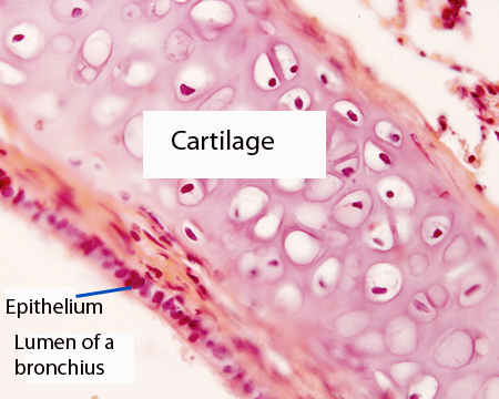
Bronchus. Staining: HE. Magnification: medium.