ELASTIC MATERIAL IN THE AORTA
The wall of the aorta as well as of the beginning of its most important branches contains a large amount of elastic material.
A cross-section of the aorta shows in its middle layer (tunica media) a large amount of parallel lines and concentricthe aortic lumen.
They stain positively for elastic material and are actually plates of elastic material whose composition is similar to that of the elastic fibers. Elastic fibers are placed in the outer region of the aortic wall.
The figures show cross sections of the plates at different magnifications.
Notice the important role of the elastic plates in the aorta:
– after each systole (contraction), the heart releases a volume of blood into the aorta.
– its wall dilates.
– after the blood has passed through each segment of the aorta, the wall returns to its previous size thanks to its elastic material, until the next systole occurs.
A certain increase in blood pressure follows each systole (the systolic blood pressure), followed by a return to the previous level (the diastolic pressure).
What would happen if the aortic wall would not go through this cycle of dilation and return to its original size?
The blood would flow as if it were inside a tube with rigid walls, such as a plastic or metal tube. The pressure in the aorta would rise significantly during systole and fall significantly during diastole. In the remaining arteries and capillaries of the whole circulatory system, the blood would advance in leaps at each systole to systole, instead of maintaining the continuous flow provided by the elasticity of wall of the aorta.
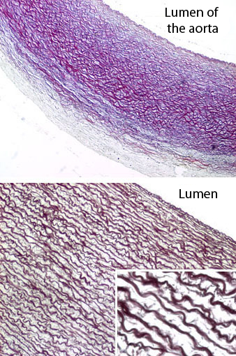
Aorta. Staining: Weigert. Low, medium, large magnifications.