RESIDENT CELLS OF THE CONNECTIVE TISSUE
MACROPHAGE-1
Macrophages are cells derived from the monocytes, a type of blood leukocyte originated in the hematopoietic bone marrow. Circulating monocytes leave the blood by crossing the walls of blood vessels and thereafter develop into macrophages in the connective tissue proper all over the body (excepting in the central nervous system), becoming concentrated in various organs such as the liver, spleen, and lymph nodes.
Being specialized in phagocytosis, the macrophages are involved in the defense of the body. Along with the neutrophils they are considered “professional phagocytes” compared to other cells that phagocyte occasionally or infrequently. Furthermore, the macrophages contains mani lysosomes and secrete several molecules that play important roles in the inflammatory process and immune response.
The macrophages are spherical or flattened, depending on the place whereh the had settled. They are relatively large cells when compared to red blood cells or lymphocytes (both measure approximately 7 µm in diameter).
The cytoplasm of the macrophages stains with eosin and its spherical nucleus is sometimes eccentric (meaning it is placed ouside of the cell center).
Recognizing a macrophage is not always easy in routine H&E-stained preparations. Immunocytochemical techniques can better identify them using specific antibodies or protein markers.
Along with the features described above, a rather good clue to identify macrophages in sections: look for phagocytosed material in the cytoplasm of isolated cells located in connective tissues or in the above mentioned organs.
The image shows liver sections of a laboratory animal that had been injected intravenously with a solution of the dye Carmine. Liver macrophages called Kupffer cells phagocytosed particles of the red dye that can be seen in their cytoplasms.
The arrows in the image indicate the phagocytosed dye. Macrophages that phagocytosed large amounts of the dye had their nuclei “pushed” toward the cell periphery.
The larger, lightly stained nuclei surrounding the macrophages belong to hepatocytes, the cells that make up the majority of the liver parenchyma. Click here if you want to recall the image of hepatocytes in Chapter 1.
Being specialized in phagocytosis, the macrophages are involved in the defense of the body. Along with the neutrophils they are considered “professional phagocytes” compared to other cells that phagocyte occasionally or infrequently. Furthermore, the macrophages contains mani lysosomes and secrete several molecules that play important roles in the inflammatory process and immune response.
The macrophages are spherical or flattened, depending on the place whereh the had settled. They are relatively large cells when compared to red blood cells or lymphocytes (both measure approximately 7 µm in diameter).
The cytoplasm of the macrophages stains with eosin and its spherical nucleus is sometimes eccentric (meaning it is placed ouside of the cell center).
Recognizing a macrophage is not always easy in routine H&E-stained preparations. Immunocytochemical techniques can better identify them using specific antibodies or protein markers.
Along with the features described above, a rather good clue to identify macrophages in sections: look for phagocytosed material in the cytoplasm of isolated cells located in connective tissues or in the above mentioned organs.
The image shows liver sections of a laboratory animal that had been injected intravenously with a solution of the dye Carmine. Liver macrophages called Kupffer cells phagocytosed particles of the red dye that can be seen in their cytoplasms.
The arrows in the image indicate the phagocytosed dye. Macrophages that phagocytosed large amounts of the dye had their nuclei “pushed” toward the cell periphery.
The larger, lightly stained nuclei surrounding the macrophages belong to hepatocytes, the cells that make up the majority of the liver parenchyma. Click here if you want to recall the image of hepatocytes in Chapter 1.
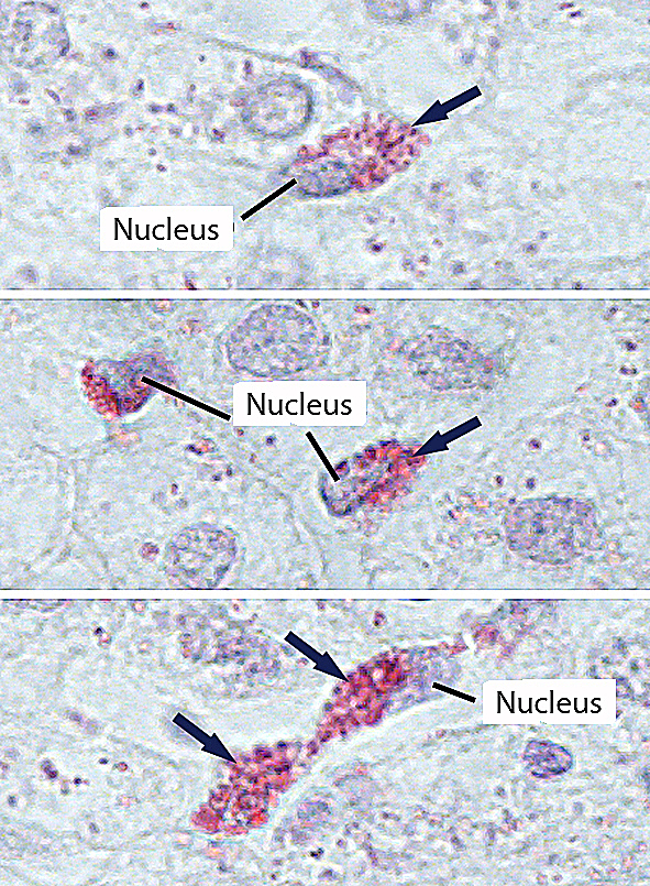
Macrophages in the liver. Staining: hematoxylin. Large magnification.