RESIDENT CELLS OF THE CONNECTIVE TISSUE – MACROPHAGE-1
The macrophage is a cell derived from the monocyte, a type of circulating leukocyte formed in the hematopoietic marrow. Monocytes leave the blood by crossing the walls of blood vessels, and settle in connective tissue, where they transform into macrophages.
Macrophages are found in all connective tissue sites. They are concentrated in various organs such as the liver, spleen, and lymph nodes, where they are involved in the body’s defenses.
Macrophages specialize in phagocytosis and, along with neutrophils, are considered “professional phagocytes,” compared to cells that phagocytize occasionally or infrequently. Furthermore, they secrete many molecules that play a role in the inflammatory process and immune responses.
The macrophage is a spherical cell and relatively large compared to a red blood cell or a lymphocyte (which measure approximately 7 µm in diameter).
Its cytoplasm is eosinophilic, stains with eosin and its spherical nucleus is sometimes eccentric, meaning it is located ouside of the center of the cell.
Recognizing a macrophage is not always easy in routine H&E-stained preparations. Immunocytochemical techniques can identify it with antibodies or proteins markers specific to macrophages.
A good way to diagnose a macrophage is to observe the presence of phagocytosed material in its cytoplasm.
The image shows the liver of a laboratory animal injected intravenously with a small solution of a dye called carmine. Liver macrophages, called Kupffer cells, phagocytosed particles of the red dye present in the blood.
The arrows indicate the phagocytosed dye. The macrophages phagocytosed a large amount of the dye, and their nuclei were “pushed” toward the cell periphery.
The larger, lightly stained nuclei surrounding the macrophages are the nuclei of hepatocytes, the cells that make up the majority of the liver parenchyma. Click here if you want to recall the image of hepatocytes in Chapter 1.
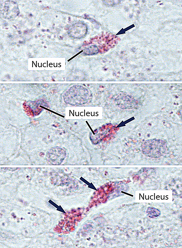
Macrophages in the liver. Staining: hematoxylin. Magnification: large.