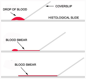
The most used way to analyze blood cells using a light microscope involves examining a blood smear, prepared as follows:
– Blood is obtained by venipuncture or by pricking the skin with a needle or stylet.
– A drop of blood is placed on a glass slide; another slide placed at the border of the drop is pulled away, dragging the blood, and producing a very thin film of blood cells.
– The film is left to dry before being stained.
Blood smears are usually stained with mixtures of dyes, called Romanovsky-type stains. There are several slightly different recipes, but almost all have in common a basic dye, an acidic dye, and a metachromatic dye.
A common composition of these solutions is a mixture of methylene blue, azure B, and eosin.
The combination of these dyes and their affinity toward cellular structures by each stain or by combinations of the dyes produce characteristic images of the various cells.
Click to review the concepts of acidic and basic dyes on this page and the following ones.
See the Glossary for the definition of metachromasia.
Important: designations of structures stained with blood dyes:
Basophilic – shades of blue.
Acidophilic, eosinophilic – pink/orange.
Metachromatic – purple.