WHITE MATTER AND GRAY MATTER
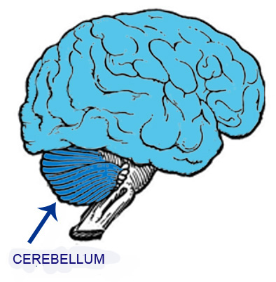
The upper figure is a schematic drawing of the brain and cerebellum, organs that will be analyzed on the following pages.
The central nervous system is formed by areas that have a high concentration of neuronal cell bodies and regions with a high concentration of neuronal extensions, mainly axons but also dendrites.
When a recently removed fresh brain or cerebellum is observed macroscopically, these two regions can be identified.
The regions with a high concentration of neuronal cell bodies have a grayish color, and are called the gray matter of the central nervous system.
On the other hand, the regions with a large number of neuronal extensions have a large amount of myelinated nerve fibers and for this reason have a whitish color. They constitute the white matter of the central nervous system.
The gray matter is an important site of reception and integration of information and responses and the white matter contains pathways between different regions of the central nervous system.
The lower figure shows the distribution of white and gray matter in the brain.
The gray matter isconcentrated in two areas:
– forming the the brain periphery, just below its surface, forming the cerebral cortex;
– in clusters of different sizes inside the brain, called brain nuclei that contain a high concentration of neuron cell bodies.
White matter occupies the interior of the brain, below the cortex and surrounding the nuclei.
The lower figure shows a cross-section of the brain and, in a simplified way, the distribution of gray and white matter in the brain.