CELLS OF THE BONE TISSUE – 3
Upper figure:
– Bone extracellular matrix: stains bluish/pink or ping after HE staining and becomes highlighted in light blue after hovering the cursor or clicking on the image.
– Osteocyte: inside a lacuna, a small cavity of the bone matrix. The osteocyte nucleus and its lacuna are highlighted in dark blue.
– Osteoblasts: active and inactive on the trabecular surface, highlighted in orange.
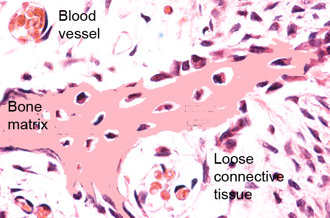
Lower figure:
Osteocytes inside a bone trabecula (highlighted in blue), surrounded by bone matrix highlighted in light blue.
Osteoblasts (highlighted in orange) on the bone surface.
Both images show active cuboidal osteoblasts and flat inactive osteoblasts or osteoprogenitors.

Bone trabecula. Staining: HE. Magnification: medium.

Bone trabecula. Staining: HE. Magnification:Medium.