HYALINE CARTILAGE – 1
Images of hyaline cartilage at different magnifications.
In hematoxylin and eosin (HE) stained sections, the extracellular matrix appears typically bluish, pale pink, or as a mixture of both colors. This staining pattern is useful for recognizing hyaline cartilage and distinguishing it from other tissues, particularly bone.
In hematoxylin and eosin (HE) stained sections, the extracellular matrix appears typically bluish, pale pink, or as a mixture of both colors. This staining pattern is useful for recognizing hyaline cartilage and distinguishing it from other tissues, particularly bone.
Upper image:: cartilage from the knee of a fetal rat. The hyaline cartilage of the bone epiphyses is highlighted in blue after placing the cursor over the image. The lighter blue band indicates hyaline cartilage of the growth plate present in long bones. The meniscus of the joint is formed by fibrous cartilage (or fibrocartilage), highlighted in dark blue.
Middle image:: tracheal wall showing hyaline cartilage. The perichondrium surrounding the cartilage is highlighted in green.
Lower image: hyaline cartilage of the wall of a bronchus.

Knee. Staining: HE. Small magnification.


Epiglottis. Staining: HE. Medium magnification.
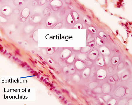
Bronchus. Staining: HE. High magnification.
NEXT PAGE