THE ARRANGEMENT OF LAMELLAR BONE IN THE DIAPHYSES – 4
The endosteum is usually a thin layer of only one cell, similar to a simple, non stratified epithelium and is in direct contact with the medullary cavity whereas the periosteum is thicker as the layer of bone cells are covered by connective tissue.
inner and
outer surfaces of the compact bone in this diagram show additional
lamellae—the outer and inner circumferential lamellae—arranged in
broad layers. The inner circumferential lamella is covered by a thin
layer of endosteum that faces the marrow cavity, similar to the outer
surface of the bone, which is covered by periosteum. Branches of
nutritional arteries accompanied by small veins are shown within the
Haversian and Volkmann’s canals. These arteries also supply the
periosteum, endosteum, and bone marrow.
interstitial lamellae They are found in the outer region of compact bone. External circumferential lamellae lie under the periosteum, while internal circumferential lamellae lie near the endosteum.
External circumferential lamellae lie under the periosteum, while internal circumferential lamellae lie near the endosteum.
Canaliculi containing the processes of osteocytes
are generally arranged in a radial pattern with respect to the
canal
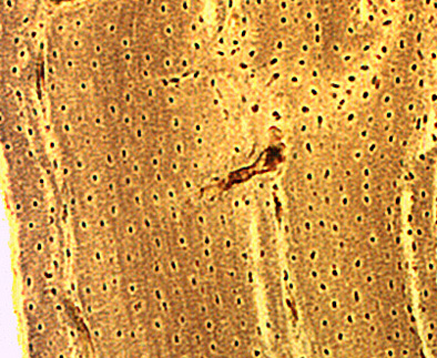
Figure 1. Outer circumferential lamellae placed under the periosteum form the outmost wall
lamellae outer circumferential system is made of stacks of slightly curved parallel bone lamellae. The lacunae occupied by osteocytes are seen as small dark circles.
Figure 2. The middle of the diaphysis wall intermediate or interstitial lamellae and Haversian systems or osteons. s. It consists of many . Between them are stretches of different lenghts of bone lamellae (highligted in red).
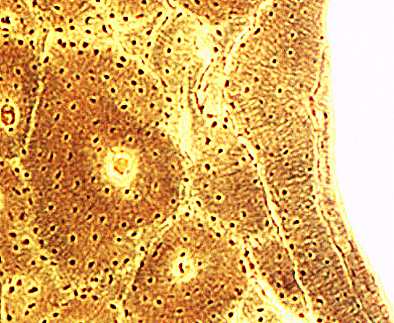
Figure 3. The internal or innner circumferential lamellae is formed by a stack of parallel bone lamellae of that covers inner surface of the diaphysis.
Haversian systems surrounded by small stretches of parallel lamellae and at the right the
1 The external or outer circumferential inner
circumferential
lamellae circumferential lamellae is made of stacks of slightly curved parallel bone lamellae. The lacunae occupied by osteocytes are seen as small dark circles.

endosteum
Elsewhere, periosteum, a fibrous connective tissue
capsule covers the outer surface of the bone.
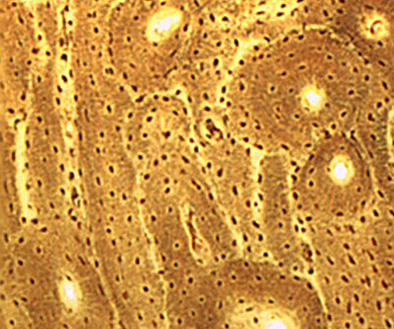
2 The central part of the diaphysis is made of stretches of different lenghts of bone lamellae (highligted in red) that constitute the intermediate or interstitial system of lamellae that placed Haversian systems or osteons. s.
It consists of many . them are
2 – The external or outer circumferential system is made of stacks of slightly curved parallel bone lamellae. The lacunae occupied by osteocytes are seen as small dark circles.
Diaphysis. aversian systems andOuter circumferential system and Haversian systems. Schmorl’s technique. Magnification: medium.
Schmorl’s technique. Medium magnification.
2 – Haversian systems and inner circumferential system. Schmorl’s technique. Magnification: medium.
Schmorl’s technique. Medium magnification.
T. Schmorl’s technique. Medium magnification.
Inner circumferential system and Haversian systems. Schmorl’s technique. Medium magnification.